EnglishFrenchGermanSpainItalianDutchRussianPortugueseJapaneseKoreanArabicChinese Simplified
If you like the site do not forget to Subscribe to our mailing list

Enter your E-mail address:

Showing posts with label CANCER. Show all posts

Wednesday, March 30, 2016

Mint can as anti-cancer drug!

Mint is one of those herbs most people associate with fresh breath or a soothing cup of tea. But researchers at India's Central Institute of Medicinal and Aromatic Plants (CIMAP) in Lucknow have been looking at it from a very different angle — as a potential weapon against cancer.

Their focus? A compound called L-Menthol, naturally found in the mint plant, which has shown a surprising ability to kill cancer cells and block their growth.

What Makes Menthol Stand Out

What makes this research particularly interesting is not just that menthol works against cancer cells — it's how it works. The compound appears to interfere with cell division, preventing cancer cells from multiplying and spreading to other organs. In laboratory studies, it has shown activity against colon cancer cells, and researchers believe it could have broader applications.

Menthol's potential isn't entirely new to science. Studies have looked at its effects on liver cancer, colon cancer, and even brain tumors. One intriguing area involves menthol-modified nanoparticles that can carry anti-cancer drugs across the blood-brain barrier — a major obstacle in treating brain tumors — achieving deeper penetration into tumor tissue than conventional formulations.

A Cost-Effective Alternative

One of the more practical arguments for exploring menthol as an anti-cancer agent is economics. Current cancer drugs are often extraordinarily expensive to produce. Paclitaxel, for instance, is derived from the bark of the European Yew tree — a slow-growing and limited resource. Menthol, by contrast, is cheap to produce, widely available, and already manufactured at scale for use in food, cosmetics, and pharmaceuticals.

That cost difference could matter enormously in making cancer treatments more accessible, especially in developing countries where high drug prices are a major barrier to care.

Research Is Still in Early Stages

It's worth being clear: this research is still primarily at the laboratory and preclinical stage. Menthol has shown genuine promise in cell studies and animal models, but it has not yet been through the rigorous clinical trials needed to confirm it as a human cancer treatment.

Scientists at the University of Salford in the UK have also been investigating a related compound, hoping to move toward testing on human cancer cells from breast and lung tissue. The path from a promising lab result to an approved drug is long and demanding — but the early signals are encouraging enough to keep researchers interested.

For now, mint remains your herb garden's most intriguing overachiever — and researchers are just beginning to understand what it might truly be capable of.


Source: Chemistry World

Tuesday, January 19, 2016

Research: Cancer Drugs may help reverse Alzheimer's disease

Alzheimer's disease and cancer don't have much in common on the surface — but at the molecular level, researchers have been finding unexpected overlaps. One of the more intriguing threads is the idea that drugs already approved to treat certain cancers might also have a role in fighting Alzheimer's.

The Connection: Tyrosine Kinases

A class of enzymes called tyrosine kinases plays a central role in cancer biology. Many cancer drugs work by inhibiting these kinases, disrupting the signaling pathways that drive uncontrolled cell growth. It turns out these same kinases are also implicated in the brain's ability to clear toxic proteins — which is exactly the process that breaks down in Alzheimer's disease.

Researchers at Georgetown University, led by Dr. Charbel Moussa, have been exploring several tyrosine kinase inhibitors for their potential to treat neurodegenerative diseases. The Georgetown group had previously shown that tau — the protein that forms toxic tangles in Alzheimer's brains — is a critical part of the cellular "garbage disposal system" that clears accumulated toxic proteins. When tau is abnormally modified (what researchers call phosphorylated tau or p-Tau), it can no longer perform this function.

The Drug: Pazopanib

One study presented at the Alzheimer's Association International Conference found that pazopanib — an FDA-approved drug for renal cell carcinoma (kidney cancer) — reduced levels of phosphorylated tau in animal models genetically engineered to overproduce human mutant tau. If that reduction in p-Tau translates to reduced toxic tangle formation, it could slow Alzheimer's progression.

This isn't the only cancer drug on the Georgetown team's radar. Their work also led to clinical trials with nilotinib, a leukemia drug, for both Parkinson's and Alzheimer's disease.

Why Repurpose Cancer Drugs?

Drug development from scratch is extraordinarily expensive and takes over a decade on average. Finding existing approved drugs that work against Alzheimer's — a strategy called drug repurposing — could dramatically shorten the timeline to treatment. Approved drugs have already cleared safety reviews, which removes one of the biggest hurdles in drug development.

The challenge is that what works in animal models doesn't always translate to humans, and Alzheimer's clinical trials have a notoriously high failure rate. But the logic of the tau pathway, and the growing evidence that tyrosine kinase inhibitors affect it, has given researchers a concrete molecular target to pursue.

The research is ongoing, and no cancer drug has yet been approved as an Alzheimer's treatment. But the intersection of cancer biology and neuroscience is producing new ideas in a field that desperately needs them.


Source: Cancer Treatment Reviews / Georgetown University

Friday, March 1, 2013

Nanotube transistors detect cancer biomarkers!!

Engineered antibodies (green/red) bound to carbon-nanotube transistors that detect osteopontin, a prostate cancer biomarker. (Courtesy: University of Pennsylvania)

What if detecting cancer could be as quick and straightforward as a routine blood test? Researchers at the University of Pennsylvania have taken a meaningful step in that direction, showing that carbon-nanotube transistors can sniff out disease biomarkers at incredibly tiny concentrations — far beyond what conventional lab methods can achieve.

The team focused on a protein called osteopontin (OPN), which is a known marker for prostate cancer and several other malignancies. By chemically bonding engineered antibody fragments directly onto carbon nanotube field-effect transistors (NT-FETs), they created biosensors that respond electrically whenever OPN binds to the antibody. In other words, the cancer biomarker essentially announces its own presence by changing how electricity flows through the nanotube device.

A Thousand Times More Sensitive

The numbers here are striking. The nanotube sensors managed to detect OPN at concentrations as low as 1 picogram per milliliter — roughly 1,000 times more sensitive than the standard ELISA immunoassay tests currently used in clinical settings. And while ELISA tests can take days to return results, these nanotube sensors deliver readings in just minutes.

Mitchell Lerner, one of the researchers on the project, put it plainly: their sensors are simpler, faster, and vastly more sensitive than existing clinical tools. That kind of leap in performance matters enormously when early detection is so critical to cancer outcomes.

How the Technology Works

The antibody fragments used are genetically engineered single-chain variable fragments (scFv) — compact, highly specific molecules that latch onto OPN with great affinity. The team attached these to the nanotube transistors using a process called diazonium salt functionalization, which creates a stable covalent bond without destroying the antibody's binding ability.

When OPN molecules in a sample bind to these antibodies, they alter the electrical properties of the nanotube — specifically, they change the current flowing through the transistor. That change becomes the detection signal. The sensors also showed strong selectivity, meaning they responded to OPN without being thrown off by other proteins in the background.

Why It Matters Beyond Prostate Cancer

Osteopontin is elevated in several types of cancer, not just prostate cancer, which means this detection approach could potentially be adapted to screen for a range of malignancies. The team also noted that the functionalization method they developed should work for any antibody with an accessible amine group — making this a platform technology, not just a one-off solution.

We're still some distance from seeing nanotube transistors in clinical labs, but the proof-of-concept is compelling. Faster, cheaper, and far more sensitive cancer detection tools could one day make early diagnosis accessible to far more patients — and that's an outcome worth working toward.


Source: Physics World

Wednesday, January 23, 2013

Flow synthesis for anticancer drug!! Amazing

UK chemists have used a combination of flow chemistry methods with solid-supported scavengers and reagents to synthesise the active pharmaceutical ingredient, imatinib, of the anticancer drug Gleevec.

The method avoids the need for any manual handling of intermediates and allows the drug to be synthesised in high purity in less than a day.

Gleevec, developed by Novartis, is a tyrosine kinase inhibitor used for the treatment of chronic myeloid leukaemia and gastrointestinal stromal tumours.

The drug molecule represents a particularly challenging target for flow chemistry because of the low solubility of many of the reaction components required for its synthesis.

The team devised a new synthesis route that prevents the equipment blockages from product precipitation and avoids many of the labour and time intensive practices of traditional batch-based preparation.
flow synthesisThe flow-based route required minimal manual intervention and was achieved despite poor solubility of many reaction components
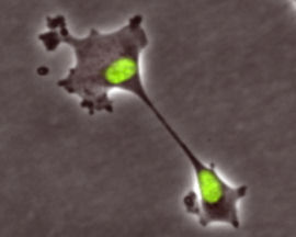
The work proved to be a challenge. Steve Ley, at the University of Cambridge, who led the team, says that along the way, they ‘met some considerable obstacles and dead ends’. He remarks that ‘in order to overcome the need to change solvents between some of the reaction stages, we had to invent a new in-line evaporator, which served us well in this and in later synthesis studies’.

Unlike the conventional industrial synthesis of Gleevec, this newly developed route couples molecular fragments in a modular approach. Thomas Wirth, who works on microreactor technology at Cardiff University, UK, remarks that ‘although not designed to compete with the industrial synthesis, the modular approach allows an easy variation of building blocks for the efficient and rapid generation of Gleevec analogues for screening purposes’.

A total of nine analogues were synthesised using the final equipment set-up, which were then screened for anticancer activity. The findings revealed that the piperazine group in the drug molecule plays a role in receptor binding, rather than simply acting as a solubilising group as previously thought.

Ley's team is now working to combine the synthesis and screening to provide information on products rapidly, as well as extending their approach to new functional materials.
RSC
------------------------- *Leave a comments, questions or even a suggestions below this post. Your expressions are always welcomed.

Monday, December 24, 2012

New type of cell division discovered (klerokinesis)!!

Klerokinesis - new type of cell division

Cell division is one of the most fundamental processes in biology — it's how organisms grow, repair tissues, and maintain themselves. We've known about mitosis and meiosis for well over a century. But a research team studying the cellular mechanics of cancer has now described a third type of cell division that nobody had previously recognized. They're calling it klerokinesis.

The discovery was made by Mark Burkard and his colleagues at the University of Wisconsin. They were investigating what happens when cell division goes wrong in ways that can lead to cancer, specifically looking at how chromosomes are distributed between daughter cells during division. What they found was a process that doesn't fit neatly into either of the known division types: in klerokinesis, the chromosomes are distributed asymmetrically in a way that appears to be partly random — more like a lottery than the precise choreography of normal mitosis.

What Makes It Different

In normal mitosis, a cell carefully duplicates all its chromosomes and then distributes exactly one complete copy to each of the two daughter cells. The process is highly regulated and essentially deterministic. Klerokinesis, by contrast, involves a more stochastic sorting of genetic material, potentially leaving daughter cells with different chromosome complements. This kind of uneven distribution is associated with chromosomal instability — a hallmark of many cancers.

Understanding this newly described process could help explain why some cells spontaneously develop abnormal chromosome numbers, a condition called aneuploidy that's common in cancer cells and is thought to drive their aggressive behavior.

What It Means for Cancer Research

If klerokinesis is a real and consistent biological phenomenon, it opens up new questions about how cancer originates at the cellular level. Could some cells be predisposed to this kind of division? Could drugs be designed to suppress it? These are early days, and the findings were presented at a scientific meeting rather than published in a peer-reviewed journal at the time of reporting, so further validation is needed. But it's a genuinely intriguing lead in the ongoing effort to understand how cancer begins.


Source: ScienceAGoGo

Friday, September 17, 2010

Scientists hail 'penicillin moment' in cancer treatment

Scientists hail 'penicillin moment' in cancer treatment
Human melanoma cells Photo: GETTY IMAGES


Scientists have hailed a 'penicillin moment' in cancer treatment following trials of a drug that uses genetic data to target the formation of specific tumours.


The study has raised hopes drug manufacturers will be able to tailor drugs to individual cancers that will halt them in their tracks and even reverse the growth of existing tumours.

The breakthrough is one of the most significant to use advances in our knowledge of DNA to tackle the root causes of disease.

For years scientists have been assembling vast amounts of genetic information provided through the human genome sequencing project. However, a number of prominent scientists had expressed disappointment that the data had not led to any major advances in treatments.

As part of the latest research, scientists in California developed a drug to block the effects of a specific gene mutation, B-RAF, linked to malignant melanoma – one of the deadliest cancers.

In one small clinical trial, tumours shrank by at least 30 per cent in 24 out of 32 patients with B-RAF mutations, and disappeared entirely in two other patients.
The drug cannot yet be declared a success: it comes with side-effects, can only treat the specific B-RAF mutation and there are no indications of its long-term usefulness.

However, a study of the chemical process behind the drug, detailed in the journal Nature, demonstrates the potential for speedy development of similar treatments targeting the particular genetic mutations that lie behind different types of tumour.

Professor Mark Stratton, Director of the Wellcome Trust Sanger Institute in Cambridge, which first linked B-RAF to malignant melanoma, said: "We've entered an end game in which we are going to complete our understanding of what causes cancer."
Yardena Samuels, a cancer geneticist at the National Human Genome Research Institute in Bethesda, Maryland, told Nature News: "It's a very important development, not just for melanoma, but for the entire cancer field."

The company behind PLX4032, Plexxikon, is now working on a test that can diagnose which malignant melanoma patients have the B-RAF mutation and would therefore benefit from the drug.
telegraph

Small dose of aspirin 'can ward off bowel cancer'

Taking a quarter of an aspirin pill every day can help prevent bowel cancer, with the benefits growing the longer it is taken, scientists claim.

A daily 75mg dose – lower than the recommended dose for a child – can provide significant protective effects after just one year, researchers said.

Even people not considered to be at high risk of the disease could benefit from taking the painkiller, they said, regardless of their diet or lifestyle.

According to the study, taking 75mg of aspirin every day for between one and three years cut the chances of developing bowel cancer by 19 per cent.

After three to five years the risk decreased by 24 per cent, and after five to ten years of taking the drug the chance of bowel cancer was down by 31 per cent.

There was no noticeable benefit in taking aspirin or any other non-steroidal anti-inflammatory drugs (NSAIDs), such as ibuprofen, for people who already had bowel cancer.
The disease, which is the second most common cause of cancer death in the world, kills more than 16,000 people in Britain every year, with 38,500 cases diagnosed in annually.

Previous research had indicated that aspirin protects against bowel cancer, but it was unclear what the most effective dose was, and how often it should be taken.
The usual aspirin dose for an adult, when used for pain relief, is between 300mg and 900mg.
NSAIDs have previously been linked to an increased risk of internal bleeding.

But the study of 2,800 people with bowel cancer and 3,000 healthy people, published in the journal Gut, showed that a much smaller dose of just 75mg could provide significant protection against the disease.
The same dose is sometimes recommended for people recovering from heart attacks and stroke.

Some 18 per cent of healthy people studied said they were taking a low dose of aspirin, or other NSAIDs, at least four times a month, along with 16 per cent of those who had bowel cancer.

While there was no evidence taking NSAIDs influenced the risk of death in bowel cancer patients, it did have a significant protective effect for those who did not have the disease at the start of the trial.

Steve Williamson, Consultant Pharmacist in Cancer at the Royal Pharmaceutical Society, said: “This study adds to the weight of evidence already around that daily low dose aspirin can reduce risk of developing bowel cancer.

"However people must remember that aspirin even at its lowest dose isn’t suitable for everyone, and patients should always talk to their doctor or pharmacist about the potential benefits of taking aspirin.”

Mark Flannagan, chief executive of Beating Bowel Cancer, said: "These findings are encouraging, particularly as, unlike previous studies, this shows that even the lowest daily dose can have an effect on risk reduction after just one year."
telegraph

Tuesday, August 31, 2010

Eight cancer signs identified!!!!!!!!

 Researchers have highlighted the eight unexplained symptoms that are most closely linked to cancer.

The Keele University team also points to the age at which patients should be most concerned by the symptoms, which include blood in urine and anaemia.

The other symptoms are: rectal blood, coughing up blood, breast lump or mass, difficulty swallowing, post-menopausal bleeding and abnormal prostate tests, reports the BBC.

Cancer Research UK said unusual changes in a person's health should be checked.

The figure for each symptom was calculated by combining the results of 25 previous studies.

They found that, if the patient was below the age of 55, there were only two signs, which reached the 'one-in-20' threshold.

These were a rectal prostate examination, which gave abnormal results, and a breast lump.

After 55, but only in men, there was evidence that difficulty swallowing could be a sign of oesophageal cancer, while blood in the urine was highlighted as a particular concern for men and women aged over 60.(ANI)

Thursday, August 5, 2010

Chemical link to testicular cancer

Teething baby
Scientists hope to prove whether common environmental chemicals, such as those used to make plastics, are to blame for rising testicular cancer rates.
Experts suspect that exposure while in the womb might explain why the rate of this cancer has doubled in 35 years.
The Edinburgh team told Human Reproduction such a study was only now possible because they had made a model to study the disease in development.
They will use mice harbouring human cells to test the theory.
Testicular cancer occurs in young men, but doctors have known for some years that the abnormal changes that lead to testicular cancer happen in the first few months that the foetus is growing.

“Start Quote

We need to know for sure if these compounds are harmful or not. The hope is that our studies can resolve this one way or another”
But because these changes occur during early pregnancy, when there is no means of studying the foetal testes, doctors do not know how and why these changes occur.
Researchers are fairly certain there must be an environmental cause because the rate of the cancer has increased so rapidly.
According to Professor Richard Sharpe, of the Medical Research Council's Human Reproductive Sciences Unit, one theory is that the changes are caused by pregnant women being exposed to environmental chemicals such as phthalates, which are used in many different household items, including plastic furniture and packaging.
But because the cancers only develop 20-40 years after the patient is born, it has been hard for doctors to discover what happened in foetal development to cause this to happen, especially when trying to establish if their mothers were exposed to phthalates or other environmental chemicals to establish a causal relationship.
Mouse model
Now the MRC researchers have developed a model in which early human foetal testis development can be studied and manipulated experimentally to establish once and for all if exposure to environmental chemicals is a likely culprit.

Related stories

Prof Sharpe's team has grafted testis tissue from aborted foetuses under the skin of mice. The germ cells in the testes are at the critical stage when any faults in their development can result in changes which make them pre-cancerous.
The researchers will expose the mice to phthalates or other environmental chemicals to see if this induces changes in the foetal germ cells that would predispose them to develop into a cancer.
Prof Sharpe said: "We are choosing to study phthalates first for several reasons, because we know that in the test-tube they can affect foetal human germ cells. They are also the most ubiquitous of environmental chemicals. We are all exposed to them."
Phthalates are used to make plastic flexible, and so can be found in carpets, wall boards, car upholstery and fittings and certain cosmetics and pharmaceutical drugs.
However, Prof Sharpe said that there was uncertainty about whether phthalate effects on the foetus in animal models were relevant to humans.
He said: "This is one of the critical unresolved questions as to whether phthalates pose a risk to human health or not.
"It's a huge industry. These compounds are literally part of the fabric of our modern society so they cannot easily be banned or removed without having widespread effects on everyday life. We need to know for sure if these compounds are harmful or not. The hope is that our studies can resolve this one way or another."
The researchers say that if phthalates do cause effects on human foetal germ cells they could know within a year. If the chemicals are not responsible it could take much longer to conclusively disprove any link.

breast cancer screening could couse health risk!!!

Mammogram
New claims suggest that breast cancer screening could be harming as many women as it helps through over diagnosis, resulting in unnecessary treatment and suffering
.


Breast cancer screening has divided the medical profession for 20 years, despite advocates of the programme claiming it annually prevents 1,400deaths and saves two women’s lives for every one who receives unnecessary treatment. 

Oxford University’s public health epidemiologist, Professor McPherson, said, ‘There is no doubt that screening for breast cancer has limited benefit and some possibility of harm for an individual woman and is of marginal cost-effectiveness for the community.’ 

Critics claim that the benefits of screening have never been properly weighed up against the drawbacks, centrally that in some cases thecancer detected does not need treatment due to slow growth or the probability that it will heal without medical interference.
A recent Danish study, published in the British Medical Journal, found that deaths from breast cancer had fallen faster in areas without screening than in those where it was used.

However, Sarah Sellars, director of the NHS Breast Screening Programme, said, ‘The vast majority of evidence from properly conducted research clearly shows that regular mammography reduces deaths from breast cancer.’

The World Health Organisation’s International Agency for Research on Cancer (IARC) concluded that there is a 35% reduction in mortality from breast cancer among regularly screened women aged 50-69 years old.

Tuesday, April 6, 2010

Tiny Drill Attacks Tough Tumors


A new approach to treating bone cancer is being tested in pet dogs.


In early February, Melissa Dauvalt noticed that her St. Bernard, Juno, had a lump on one of his front legs that was causing him to limp.


Tests confirmed that the dog had developed osteosarcoma, an aggressive and difficult-to-treat bone tumor that's found most commonly in adolescent children and large-breed dogs.

Dauvalt is a veterinarian, so she knew the most effective treatment would be amputation.

But she couldn't bear the thought of her 165-pound canine trying to walk on three legs.

"He's a big guy," says Dauvalt. "Amputation was not an option."

So Dauvalt and volunteered her dog for a clinical trial at Texas A&M University, where veterinarians are testing a new technology that delivers radiation directly to solid bone tumors. During a two-hour procedure on March 12, the vets used a drill roughly the size of an electric toothbrush to inject a radioactive isotope directly into the tumor, in the hopes of shrinking it without harming surrounding tissue.

The lessons they learn from Juno's experience--and that of other dogs in the trial--will help scientists determine if such highly targeted radiation treatments could also work in humans.

Osteosarcoma afflicts about 900 people per year in the United States, about half of whom are younger than 20, according to the American Cancer Society.

Some tumors can be removed effectively, but what makes the disease so devastating is its tendency to spread: One in four newly diagnosed cases have already metastasized, and the long-term survival rate is under 25 percent, says Crystal Mackall, chief of the pediatric oncology unit of the National Cancer Institute.

Osteosarcoma is genetically similar in dogs and humans, making the veterinary setting an important laboratory for researching human therapies.
"Dogs develop cancer naturally, and they share our environment," says Melissa Paoloni, a scientist at the NCI's Comparative Oncology Program.

That provides an opportunity to try potential new treatments in something other than rodents, which have to be engineered to develop cancer and therefore may not most accurately reflect the human experience.


About 6,000 new cases of canine osteosarcoma are diagnosed each year, according to the National Canine Cancer Foundation.

The drill was the brainchild of Stan Stearns, founder and president of Valco Instruments, a Houston-based maker of medical devices. Stearns lost his St. Bernard, Gabriel, to osteosarcoma in 2008. During the yearlong effort to save Gabriel's life, Stearns learned that external-beam radiation--the type commonly used to treat cancer--wasn't really effective for bone tumors, and presented too much of a risk to surrounding healthy tissues.

But after reading about experiments using biopsy tools to deliver medicine directly to bone, he had an epiphany.

"Valco had developed tubing that I thought could be the basis of a drill that would be flexible enough to go around corners," burrow into bone, and deliver radiation directly to tumors, Stearns says.

Valco's engineers developed the drill in a matter of months, and then partnered with startupIsoTherapeutics in nearby Angleton, TX, to formulate the radioactive solution.

IsoTherapeutics was founded in 2005 by two veterans of Dow Chemical who set out to develop "radiopharmaceuticals"--radioactive compounds designed to shrink tumors. They combined a radioisotope called yttrium-90 with a proprietary formula that prevents the compound from traveling beyond the tumor. The drill makes several tiny holes in the bone surrounding the tumor, and then delivers microscopic amounts of the compound into each hole. Because yttrium-90 emits high-energy beta particles, "we can cover the whole tumor with a minimal dose," says Keith Frank, cofounder of IsoTherapeutics.

And yttrium-90 does not emit gamma radiation, which greatly reduces the risk of radiation exposure to people working with the compound.

The Texas A&M veterinarians who performed Juno's procedure were guided by a high-powered PET/CT scanner. "We were able to see a perfect outline of the leg," Fossum says. After the team injected a contrast agent that binds to glucose in tumors, the PET/CT "showed us where to inject the radioisotope," says Theresa Fossum, a professor of veterinary surgery at the university.

Sixteen dogs have been treated with the experimental procedure at a handful of veterinary schools so far, Frank says. While it's too early to quantify the results, he says, "we always see pain palliation, and we definitely see an increase in survival

" He believes the procedure will work on other types of bone tumors, too.

IsoTherapeutics is currently in talks with researchers who want to test the technique in women with breast cancer that has metastasized to bone, Frank says.

As for Juno, he was walking without a limp 10 days after his treatment.
He'll undergo chemotherapy to attack any latent cancer cells, and then have a follow-up scan in about six months.

Owner Dauvalt says she has no regrets, regardless of what the outcome might be.

"We have to find different ways of treating cancer," she says. "I felt this was an opportunity not only to help my dog, but possibly to help people."
technologyreview